In Turkey, Milia is the term used to describe Milialar. This term refers to the small cysts, which look like pearls, that are commonly found on the skin surrounding the eyes and on the eyelids. These small bumps, although generally harmless in nature, can cause distress due to their appearance. Note that milialar does not fall under the category of acne. This comprehensive guide will explore the world of milialar, or milia. We’ll look at its types, symptoms, signs, prevention measures, and treatment options. This information is based on scientific research and expert insight on milia. It will help you to understand this condition.
What is Milia, or Milialar
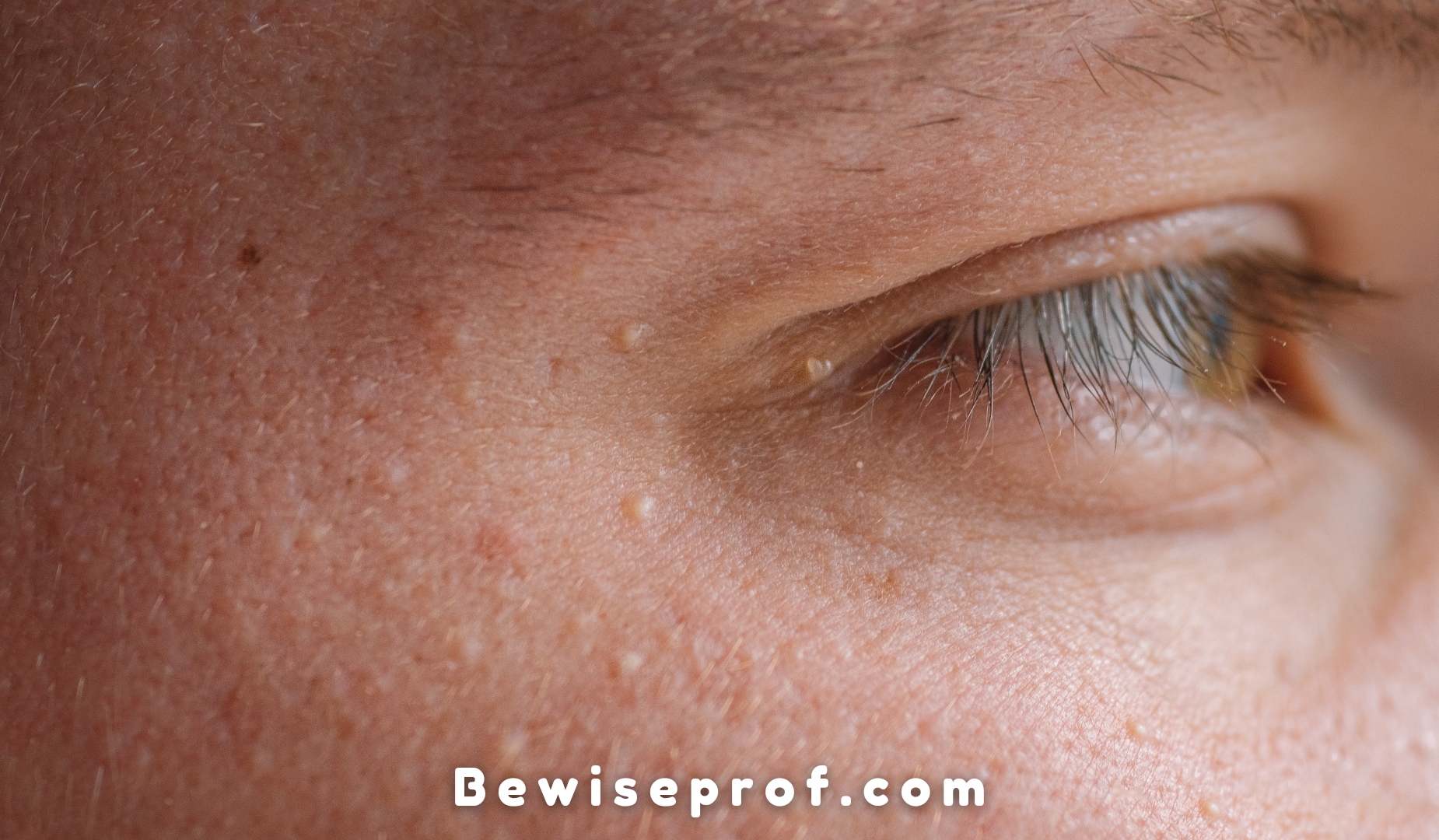
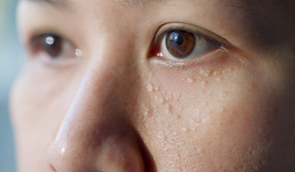
Milialars are small, dome-shaped bumps with a size between 1-2 millimeters. They’re about the same size as a pinhead. The cysts are pearly white with a smooth and firm texture. The most common place to find milia is on the eyelids and under the eyes. They look like tiny pearls. A study found that milialar is caused by keratin (a protein in skin, and hair) becoming trapped under the surface of the skin. Adults can develop milialar as well, but they’re more common in newborns.
Fun fact Despite their similar sounding names, milia or milialar is not related to malaria. Malaria is caused by parasites.
Milia or Milialar: Types and Categories
There are two types of Milialar: primary Milialar, and secondary Milialar.
Primary Milia or milialar
The primary milia is formed by the direct entrapment keratin in the skin. These are more common in neonates, due to the immature sweat glands. The following are the key characteristics of a primary milia.
- Small, white-to-yellow cysts.
- Commonly found around the eyes, nose and cheeks.
- Asymptomatic.
- In infants, the condition usually resolves on its own within a couple of weeks or months.
Second Milia or Milialar
The secondary milia are caused by trauma to the or skin. Adults can develop them after certain skin conditions and procedures. Secondary milias or milialars have the following characteristics:
- Similar appearance to primary milialar. Often seen where a procedure or injury has occurred.
- The underlying cause of the symptoms, like pain after a burn, may be causing them.
- The duration of the condition can vary and last longer, depending on its cause.
- Treatments such as laser therapy, manual extraction or medication may be used to address the underlying causes.
What causes milia or Milialar development?
Milialar cysts form when dead cells are trapped beneath the surface of the skin. They can appear anywhere on the body, but they are most common on the face. The development of milia and milialar is influenced by several factors, but the trigger may not be always known. These include:
- Genetics Some people have a predisposition to develop Milia. This is often inherited.
- Sun Exposition: Prolonged exposure to the sun can cause facial skin damage over time and increase the risk of developing milia.
- Skin trauma: Injuries to the skin such as burns, blisters, abrasions and cuts can lead to milia.
- Some Medical Conditions Disorders that cause inflammation and dry skin, such as eczema can increase your risk.
- Medicines Some medications like steroids may cause milia.
- Heavy Makeup and Creams: Use of thick, greasy products may cause cysts.
The most common milia is found in newborns. Up to 50% of babies develop transient milia, which usually disappears within a couple weeks. This is thought to be due to maternal hormones. Persistent milia affects 2.5% of adults. Women are affected more than men and milia becomes more common with age. It is thought that aging-related changes to skin cell kinetics, as well as decreased skin elasticity, cause milia.
Development Process
The process of developing milialar is very specific:
- Skin Regeneration: The skin sheds dead cells as part of its natural renewal process. These cells can sometimes fail to shed correctly.
- Trapped Keratin : The cells trapped then form keratin which accumulates.
- Cyst Formation: The accumulation of milia leads to the formation of small cysts under the skin.
Expert insight: Although there are many subtypes of Miliar, the most common are those that occur spontaneously around the eyelids due to keratin accumulation. Secondary milialar may be caused by trauma, blistering, burns or other ophthalmic conditions.
Milia and Milialar: Symptoms and signs
Milialar can be easily identified by their distinctive appearance. They can manifest themselves as:
- The eyelids and around the eye may have small, pearly-white bumps.
- Under the skin, you will find smooth bumps that look like pearls.
- Yellowish-white or whitish-yellow in color.
- Can appear in single or clusters.
- They are usually painless, and do not cause irritation or itching.
- They can remain unchanged for several weeks or even months, or they may disappear by themselves.
- If ruptured, the syringe may release a cheese-like waxy discharge.
Tip : Consult a dermatologist if you are unsure about a skin condition.
Preventive measures
Although it is not possible to completely prevent milialar, following these tips can help reduce the risk.
- Use makeup and moisturizers that are non-comedogenic, oil-free.
- Avoid using heavy creams or cosmetics around the eyes.
- Exfoliate your skin and cleanse it gently to remove clogged pores.
- Use the correct technique to shave your skin.
- Use sunscreen every day and limit your sun exposure.
- To prevent excessive dryness, keep your skin hydrated.
- Before bed, remove all makeup and old makeup.
- Treatment of underlying skin conditions such as eczema.
- Avoid intensive facials and chemical peels if you have milia (also called millia). These can worsen the condition.
Treatment options for Milia or Milialar
Most milialar bumps do not need treatment and most resolve on their own within a few weeks or months. If the bumps are persistent or cause discomfort, there are several options for treatment:
- Retinoid Creams Creams that contain tretinoin or adapalene can help to dry and remove the milia.
- Microdermabrasion This technique uses fine crystals that gently exfoliate and stimulate the healing of outer skin layers.
- Chemical peels: A mild solution of glycolic or salicylic acids can be applied to soften the lesions and remove them.
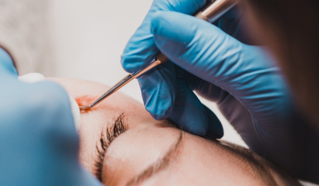
- Electrocautery involves the burning of the milialar using a hyfrecator cauterizing tool, while using a local anesthetic.
- Manual Removal A dermatologist may open the cyst using a sterile syringe and squeeze the contents out.
- Cryotherapy : freezing the bumps using liquid nitrogen in order to eliminate lesions.
- Laser ablation: Use laser energy to destroy cysts.
- Surgical Removing: A dermatologist might opt to remove milias surgically by opening them and draining out the fluid. Sometimes, stitches are required.
Important Notice This information is for educational purposes only. It is recommended that you seek professional advice before beginning any treatment.
The conclusion of the article is:
The small cysts, which look like pearls, that appear on the skin are usually harmless. They can occur on the eyelids or around the eyes. Adults can develop them as well, but they are usually more common among newborns. This is often due to skin damage. Milialar is found in many types and its development can be influenced by a variety of factors including genetics.
There are many treatment options for milia, but most cases will resolve themselves. It is important to practice good skincare, avoid using heavy cosmetics and no make-up, as well as protect your skin against excessive sun exposure.
This guide will help you understand milia in detail, including its types, symptoms, prevention measures, and treatment options. Consult a dermatologist if you are concerned about the condition of your skin or milia.